Intraoral Photos Salem, MA
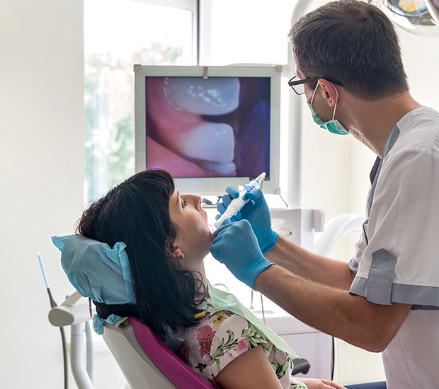
Intraoral cameras allow dentists to expand their interpretation and perception of a patient's condition. They also assist in the execution of a well-made, personalized treatment plan. Intraoral photos may reveal any defects in the teeth and other parts of the oral cavity that may have otherwise gone unnoticed.
Dental photos are available at Burba Dental Partners in Salem and the surrounding area. Intraoral photography can help enhance patient education and expedite the healing process. Call us today at (978) 703-2008 to schedule an appointment or to learn more about our services.
The History of Intraoral Cameras
The first intraoral camera was developed in 1987. These cameras were large, unwieldy, and pricey, at an average cost of about $40,000 per unit, and they consisted of a dental endoscope, a light scope, and a remote head micro camera. They required a handpiece, video processor, and dedicated computer to process the captured images and videos.
Today's intraoral camera systems are much more portable and easy to use. They have USB connectivity instead of a large docking station, making them lightweight and ergonomic. Many look just as conventional cameras do, and they can produce higher-quality images and videos that are readily available to view. As such, they have become so easy to use that they are quickly becoming a standard part of many dentists' operatories.
“Today’s intraoral camera systems are much more portable and easy to use.”
When to Use Intraoral Cameras
Intraoral cameras are an invaluable diagnostic tool. They facilitate patient education and help dentists better communicate with their patients. They are used regularly in dental examinations, particularly when determining accurate charting, assessing caries risk, and identifying restorative materials (both existing and ideal). Intraoral cameras are frequently used in place of mirrors, probes, and radiographs to evaluate the teeth.
Furthermore, intraoral cameras can help dentists show the importance of oral hygiene. By allowing patients to visualize the state of their oral health, dentists can use intraoral cameras to demonstrate proper oral hygiene practices. This may also enable patients to better understand any oral health conditions they may be afflicted with and, thus, assist in informed decision-making in the future.
“…intraoral cameras are frequently used in place of mirrors, probes, and radiographs to evaluate the teeth.”
What Intraoral Photos Reveal
Since their inception in the 1980s, intraoral cameras have continually helped dentists make more accurate diagnoses, establish a foundation of trust for relationships with patients, and determine the best treatment course for each patient's unique condition. They are particularly apt at detecting occlusal cavities, especially in comparison to visual examinations and microscopes.
Dental photos can also help establish a baseline for any existing dental conditions. This makes it easier for dentists to evaluate the mouth for any suspicious lesions, recession, or other aberrations. They may include the teeth, gums, or oral tissue. It is easier to see when an amalgam filling needs to be replaced, such as when dental photos make existing stress lines, wear, and discoloration evident.
“Dental photos can also help establish a baseline for any existing dental conditions.”
Check out what others are saying about our dental services on Yelp: Intraoral Photos in Salem, MA
Intraoral Photos and Diagnosis
Even today, it is not uncommon for some dentists to diagnose certain conditions after an unassisted visual examination. Though these may sometimes suffice, a more in-depth inspection is occasionally necessary. Intraoral cameras allow dentists to take close-up photos of the teeth from angles that they could not otherwise see, facilitating the diagnosis process. This can even be true when a patient has undergone an X-ray, as certain conditions may be more apparent "in the flesh."
Taking dental photos regularly also allows dentists to establish a "baseline" for a patient's mouth. Any aberrations from these photos will then be more easily identifiable as potential causes for concern. Dental photos can also make visits more convenient for the patients, who may have otherwise needed to be subjected to hours of visual examination for a proper diagnosis.
“Intraoral cameras allow dentists to take close-up photos of the teeth from angles that they could not otherwise see, facilitating the diagnosis process.”
Questions Answered on This Page
Q. What is an intraoral camera?
Q. What are intraoral cameras for?
Q. How can intraoral cameras assist in my treatment?
People Also Ask
Q. What does the dentist look for in a dental examination?
Q. Why is preventative care important? How can it save you money?
Frequently Asked Questions
Q. Will my insurance carrier accept dental photos as verification of my condition?
A. The answer depends on your carrier and condition. However, most insurance carriers require some form of charting, radiographs, or narrative before disbursing benefits. Intraoral photographs can often help to support a patient's case.
Q. How are dental photos useful in prosthesis?
A. Stone models are commonly used to depict a patient's tooth position and shape. Unfortunately, these models fail to capture tooth or gingival character, color, or shade. Color-corrected intraoral photos can assist in creating the most natural-looking prosthetic teeth for the patient.
Q. What should I expect when my dental photos are being taken?
A. We may take intraoral photos before, during, or after your treatment. In any case, you will need to rest comfortably in one of our chairs and hold certain positions. We will set up lights and mirrors to get the most accurate depiction of the mouth, and we may hold back your lips as necessary to allow for a good view of the mouth and teeth. Once we have taken the photographs, Burba Dental Partners will review them with you and point out both the healthy areas and potential areas of concern.
Q. What are some dental conditions that you can identify with intraoral photos?
A. There is a wide array of health conditions we may be able to detect with intraoral photography. Some of the most common are cavities, cracks, decay, enamel texture, gum disease, and tooth and gum abnormalities.
Q. Do I need to take intraoral photos every time I go to the dentist?
A. No. Every patient has different needs. Some may need to take more frequent intraoral photos, and some may need to take less. In any case, they are generally not required during every check-up. Burba Dental Partners may use intraoral photography to establish any existing conditions or make a diagnosis before creating a customized treatment plan for you.
Dental Terminology
Call Us Today
Dental photography can help patients better understand their conditions and maintain their oral health. We at Burba Dental Partners can help. Call us today at 978-703-2008 to schedule an appointment or to learn more about our services.
Helpful Related Links
- American Dental Association (ADA). Glossary of Dental Clinical Terms. 2024
About our business and website security
- Burba Dental Partners was established in 1969.
- We accept the following payment methods: American Express, Cash, Check, Discover, MasterCard, and Visa
- We serve patients from the following counties: Essex County, Middlesex County and Suffolk County
- We serve patients from the following cities: Salem, Peabody, Danvers, Beverly, Marblehead, Swampscott, Lynn, Lynnfield, Wakefield, Saugus and Boston
- National Provider Identifier Database (1952330516). View NPI Registry Information
- Healthgrades. View Background Information and Reviews
- Norton Safe Web. View Details
- Trend Micro Site Safety Center. View Details
Back to top of Intraoral Photos
